

MEDICAL LEGAL: UTV ROLLOVER ACCIDENT
Open Reduction and Internal Fixation (ORIF) of the Right Olecranon Fracture
…………….

STORY
…………….
This piece is part of a series of six medical legal visualizations, designed to depict a legal case involving a UTV rollover accident. In this incident, a 57-year-old woman was ejected and suffered substantial injuries to her right arm. The challenge of this project was to create an exhibit of illustrative evidence to clarify complex medical and scientific concepts to a courtroom audience. Moreover, maintaining adherence to style guides and meeting client expectations was crucial for creating a cohesive series of medical visualizations. This particular exhibit focuses on illustrating the open reduction and internal fixation of an olecranon fracture sustained during the rollover.
Audience: General public, jurors
Software: Photoshop, Procreate, Figma
Format: Courtroom presentation, Print
Client: Stephen Mader, University of Toronto
Key Skills: reading radiographic data and medical files, gross anatomy knowledge, science communication to lay audiences, visual storytelling, digital painting, layout
DETAILS
…………….
PRE-PRODUCTION
…………….
CASE DESCRIPTION
This project began with reading through the client’s medical documents in order to extract details of the injuries she sustained and subsequent surgeries and complications she experienced. The following is a summary of this research:
The client is a 57-year-old woman who was a passenger in an UTV rollover accident. She was wearing a helmet, but not seatbelt, and was subsequently ejected from the vehicle.
Injuries sustained:
Right hand avulsions and lacerations
Comminuted fracture of the right distal radius
Comminuted fracture of the right olecranon process
Proposed Exhibits
Hand laceration
Right olecranon fracture
Right distal radius fracture
Complications
Right ulnar osteotomy
Right extensor carpi ulnaris (ECU) release
Surgery:
Open reduction, internal fixation of right distal radius fracture
Open reduction, internal fixation of olecranon fracture
…………….……….……….
…………….
DRAFT ILLUSTRATION
Complications:
Malunion
Osteoarthritis
Osteopenia
Pain
Corrective Surgery:
Right ulna shortening osteotomy
Right extensor carpi ulnaris release

This exhibit focuses on the fracture to the right olecranon process caused by the accident. The orientation image demonstrates the position of the arm with respect to the viewer. Radiographs and an illustration of the injury are depicted on the left, followed by a larger post-op illustration of the open reduction internal fixation (ORIF) surgery along with radiographs.
Details of Injury:
Comminuted fracture of the olecranon process; two osteochondral fractures.
Details of surgery:
A metal compression plate was placed across the comminuted fracture. Four screws were inserted distally, four were inserted proximally. A local bone graft was used to repair and rebuild the damaged bone.
…………….
FINAL ILLUSTRATION
…………….
STYLE GUIDE

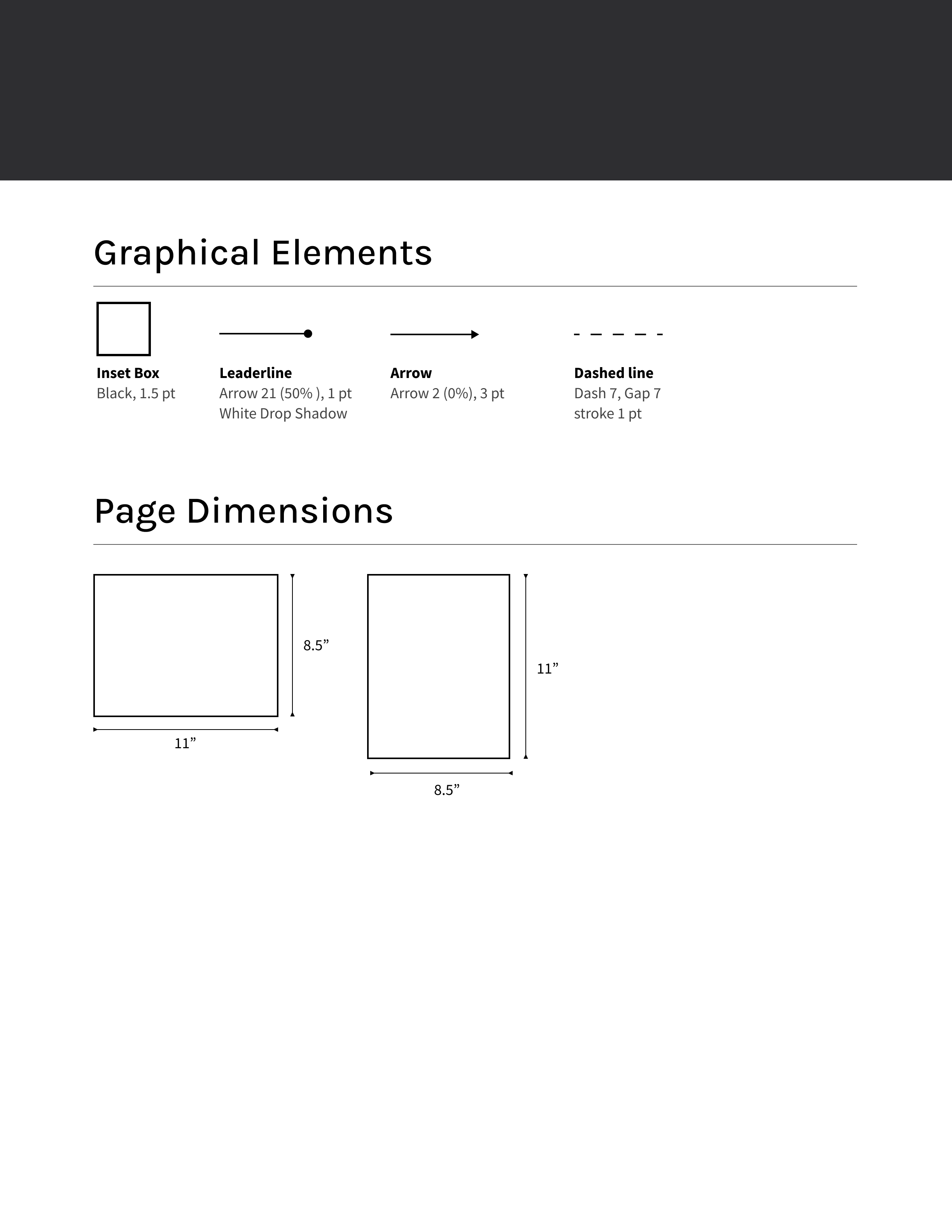
In reality, anatomical structures that are viewed mid-surgery are red because of the abundance of blood. For a didactic illustration, the following was considered:
Colors were chosen so that different anatomical structures can be easily identified by color (adipose in comparison to bone).
Anatomical conventions of color were followed so viewers can apply their knowledge of anatomy that they have seen elsewhere (i.e: adipose tissue is an exaggerated yellow color).
As opposed to a purely didactic illustration, less saturated colors were used to better reflect the serious tone of the patient's medical trauma